
The scalp, though often hidden beneath hair, is a vital part of our overall health. It plays a crucial role in hair growth and can be susceptible to various conditions. In scalp dermatology, accurate diagnosis is key to effective treatment. This is where digital microscopes come in, offering a powerful tool for magnified examination and improved patient care.
Traditional Challenges in Scalp Examination
Traditionally, scalp evaluation relied on the dermatologist’s naked eye and handheld magnifying devices. While these methods provide a basic assessment, they have limitations. Visualizing subtle details like hair follicle miniaturization (follicles shrinking) or specific types of scaling can be difficult. Additionally, documenting findings for comparison over time or sharing with colleagues can be challenging.
Using Dino-Lite to Assess Scalp Health
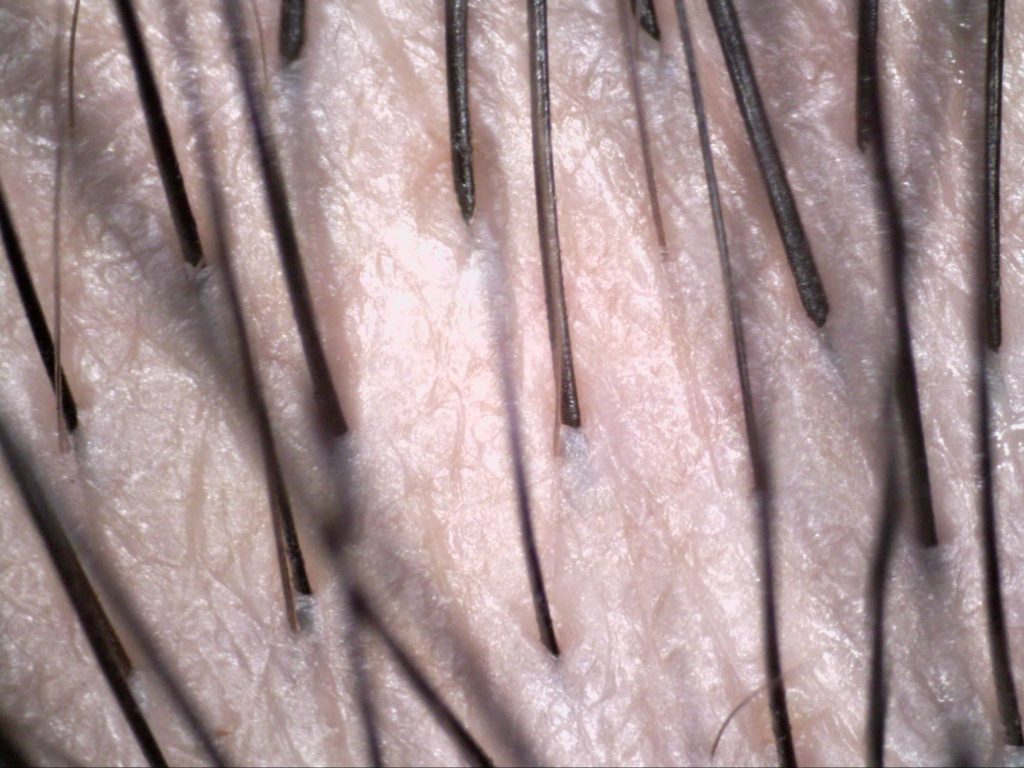
Digital microscopes bridge this gap by offering high-resolution magnification, typically ranging from 20x to 200x or even higher. This allows dermatologists to see intricate details of the scalp, including:
- Hair Follicle Characteristics: Assess the size, shape, and density of hair follicles to diagnose conditions like androgenetic alopecia (pattern hair loss) or alopecia areata (autoimmune hair loss).
- Scalp Conditions: Identify and differentiate between various scalp issues like seborrheic dermatitis (dandruff), scalp psoriasis, and folliculitis (inflammation of hair follicles).
- Parasites and Microorganisms: Detect the presence of lice, mites, or fungal infections on the scalp.
Benefits of Digital Microscopy in Scalp Dermatology
The use of digital microscopes in scalp dermatology offers several advantages:
- Improved Diagnostic Accuracy: High-resolution visualization allows for more precise diagnosis of scalp conditions, leading to better treatment decisions.
- Enhanced Documentation: Captured images and videos provide valuable documentation for patient records, enabling better tracking of treatment progress and comparison over time.
- Teledermatology: Digital images facilitate teledermatology consultations, allowing remote evaluation of scalp conditions and potentially improving access to specialist care.
- Patient Education: By visualizing the magnified view of their scalp, patients can gain a deeper understanding of their condition and treatment rationale.
The Future of Scalp Dermatology with Digital Technology
Digital microscopy is a rapidly evolving field with ongoing advancements. Integration of artificial intelligence (AI) for automated image analysis is a promising area of development. AI algorithms can potentially assist in identifying specific scalp conditions or measuring hair follicle parameters, further streamlining diagnosis and treatment planning.
In conclusion, digital microscopes are revolutionizing scalp dermatology by offering a magnified view of scalp health. Their ability to provide high-resolution images, improved documentation, and potential for AI integration makes them a valuable tool for accurate diagnosis, effective treatment, and enhanced patient care. As technology continues to advance, digital microscopes are poised to play an even greater role in the future of scalp dermatology.
