
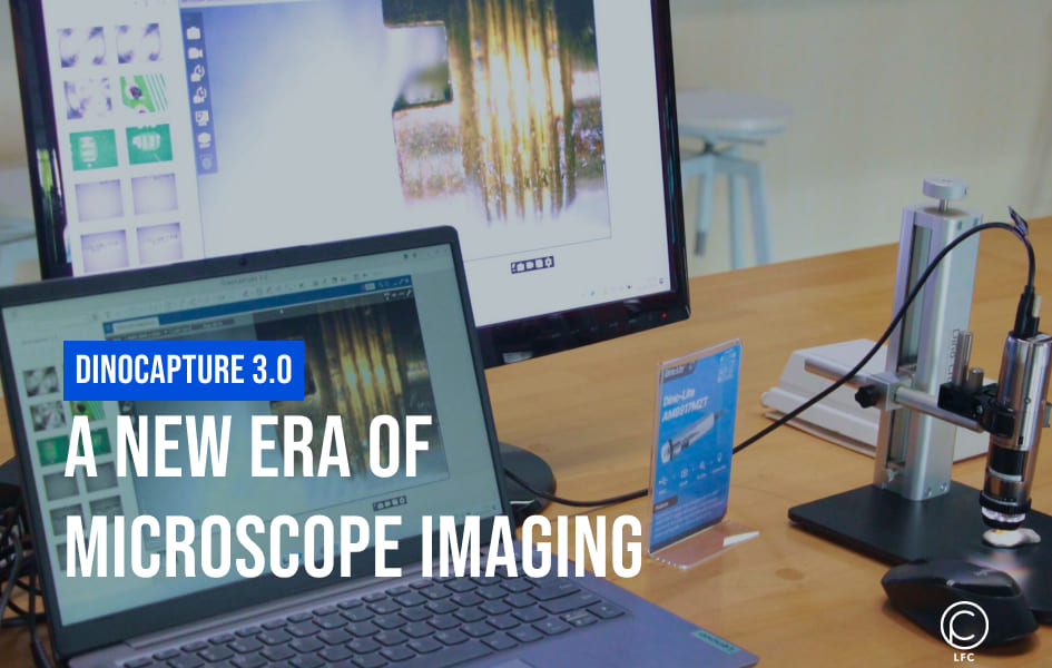
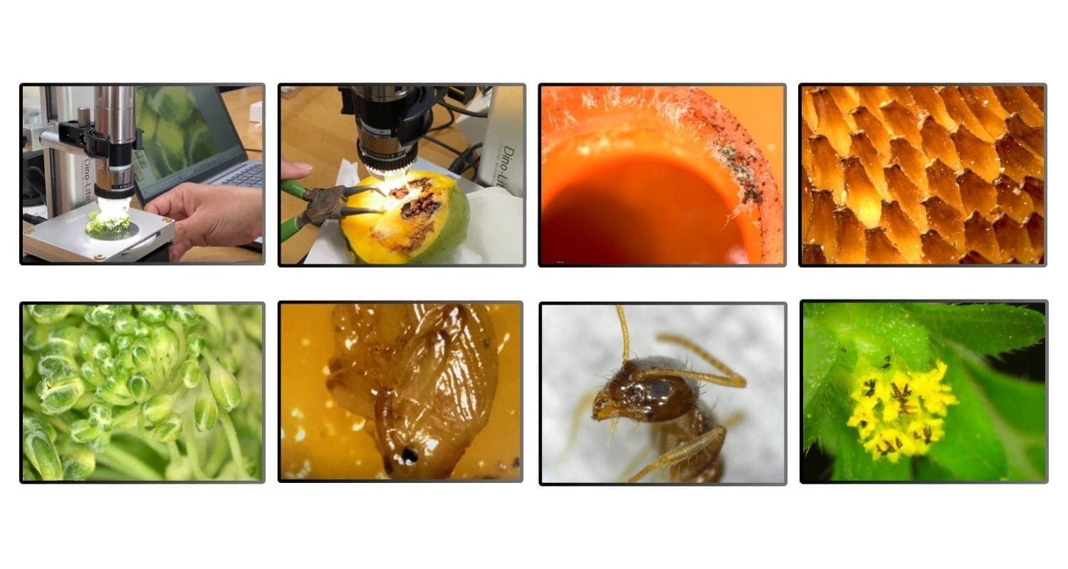
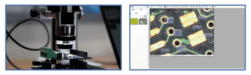
DinoCapture 3.0: Unlocking the Microscopic World
Dino-Lite, a leader in digital microscopy, has introduced DinoCapture 3.0, a major update to their well-regarded microscope imaging software. This robust software is engineered to optimize your microscopy experience, providing tools to improve image clarity, measurement precision, and workflow productivity.
DinoCapture 3.0 version 1.1.0.0 is available for download (click here).
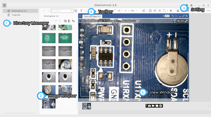
Key Features of DinoCapture 3.0
1. Enhanced Performance
- Experience a significant performance boost.
- Capture MP4 videos for versatile use and smaller file sizes.
- Simultaneously capture images, videos, EDOF, and EDR on multiple cameras.
2. Improved File Structure
- Embedded data for enhanced portability.
- Utilise the file explorer for easy search, rename, sort, move, and favourite functions.
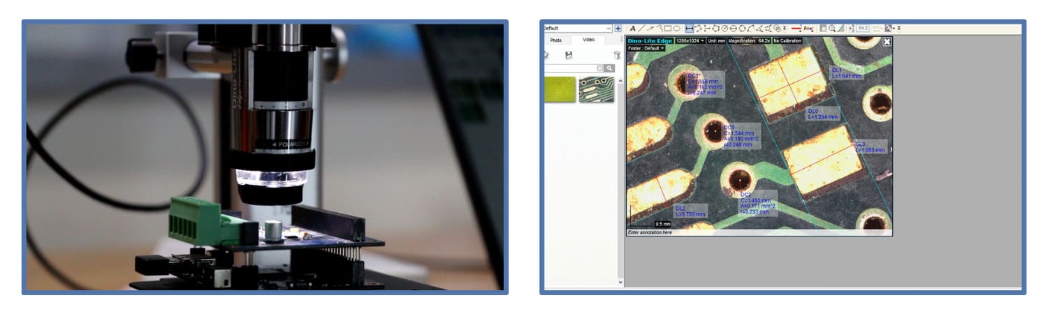
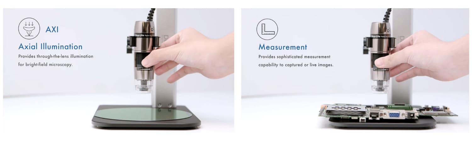
3. Enhanced Measurement
- Subpixel coordination for more accurate measurements and drawings.
- ROI measurement for precise results, especially for repetitive tasks.
- Reduced measurement variability with the ROI measurement tool.
- Measurements were available in Line, Distance, Circle, Arc, Angle, Chamfer, and Roundover.
- Collect measurement data in a lightweight database for efficient management.
4. Compatibility
- Dino-Lite and Dino-Eye USB models: Legacy models requiring driver installation are not supported.
- Operating System: Windows 10/11
Benefits of DinoCapture 3.0
Here are the key benefits derived from the enhanced features of DinoCapture 3.0:
- Enhanced Performance: Faster processing, smoother operation.
- Advanced Measurement: Precise measurements and data analysis.
- Intuitive Interface: User-friendly design for easy navigation.
- Versatile Applications: Suitable for various microscopy tasks.
- Efficient Workflow: Streamlined processes for increased productivity.
Elevate Your Microscopy Experience
By leveraging these powerful features, DinoCapture 3.0 empowers users to explore the microscopic world with unprecedented detail and precision. Whether you are a researcher, educator, or hobbyist, DinoCapture 3.0 is the ideal tool to enhance your microscopy experience.
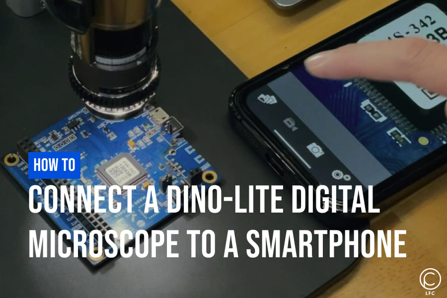
Step-by-Step Guide: Connecting Your Dino-Lite Microscope to a Smartphone
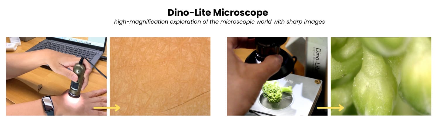
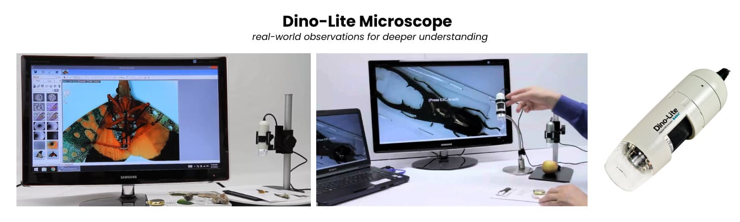
A Dino-Lite digital microscope is a compact, portable, and versatile handheld device designed to perform many of the functions of a traditional optical microscope. Despite its small size, it delivers powerful magnification and imaging capabilities, making it ideal for various applications, from scientific research and education to industrial quality control and electronics inspection. Equipped with advanced features such as high-resolution imaging, adjustable magnification, and specialized lighting options, the Dino-Lite offers exceptional convenience and flexibility. Its portability allows users to conduct detailed inspections and analyses in various settings, whether in the lab, in the field, or at a workstation, making it a valuable tool for professionals and hobbyists.
Why Use Dino-Lite WF-10 and WF-20 Adapters?
When used with Dino-Lite digital microscopes, WF-10 and WF-20 adapters are primarily designed to enable wireless connectivity with smartphones, tablets, or computers. While both adapters serve the same purpose, they may differ slightly in features and performance.
 |
 |
| Dino-Lite WF-10 Adapter | Dino-Lite WF-20 Adapter |
Choosing the Right Adapter for Smartphone Connection
1. WF-10
-
Compatible with most Dino-Lite microscopes with USB interface.
- It can be powered by a power adapter or a battery, offering flexibility in usage scenarios.

2. WF-20
-
Compatible with every Dino-Lite AF-series model.
-
Provides over 2.5 hours of battery life, allowing for extended usage without needing a power source.

Note: The WF-10 or WF-20 Wi-Fi streamer is compatible with the DinoConnect app for iOS and Android devices. For Windows users, it works with DinoCapture 2.0 version 1.5.28 or later.
Connect a Dino-Lite Microscope to a Smartphone Using WF-10
1. Download the DinoConnect App
-
Download the free DinoConnect app from the App Store (iOS) or Google Play Store (Android).

2. Connect the Adapter and Power On
-
Connect the Dino-Lite microscope to the labelled USB port on the WF-10 adapter.
-
Power on the WF-10 adapter.
3. Connect to the Wi-Fi Network
-
Turn on Wi-Fi on your smartphone or tablet.
-
Connect to the Wi-Fi network named "Dino-Lite WiFi Streamer."
-
The default password is 12345678.
 |
 |
4. Open the DinoConnect App
-
Launch the DinoConnect app on your device.
5. View and Control
-
The app will display a live feed from the microscope on your device's screen.
-
You can capture images, record videos, and adjust settings directly from the app.

Connect a Dino-Lite Microscope to a Smartphone Using WF-20
1. Download the DinoConnect App
-
Download the free DinoConnect app from the App Store (iOS) or Google Play Store (Android).
2. Attach the WF-20 Wi-Fi Streamer
-
Remove the USB adapter from your Dino-Lite microscope.
-
Attach the WF-20 Wi-Fi Streamer to the microscope.
-
Power on the WF-20 Wi-Fi Streamer.
 |
 |
3. Connect to the Wi-Fi Network:
-
Turn on Wi-Fi on your smartphone or tablet.
-
Connect to the Wi-Fi network named "Dino-Lite WF-20."
-
The default password is 12345678.
4. Open the DinoConnect App
-
Launch the DinoConnect app on your device.
5. View and Control
-
Your smartphone's screen will display a live view from the microscope.
-
You can capture images, record videos, and adjust settings directly from the app.
Dino-Lite Distributor in Singapore
LFC PTE LTD is the authorized distributor of Dino-Lite in Singapore. Our expert team can assist you in selecting the ideal microscope to meet your specific requirements.
Follow LFC PTE LTD on social media to stay updated on Dino-Lite products and other visual equipment. You can find us on Facebook, Instagram, YouTube, and LinkedIn.
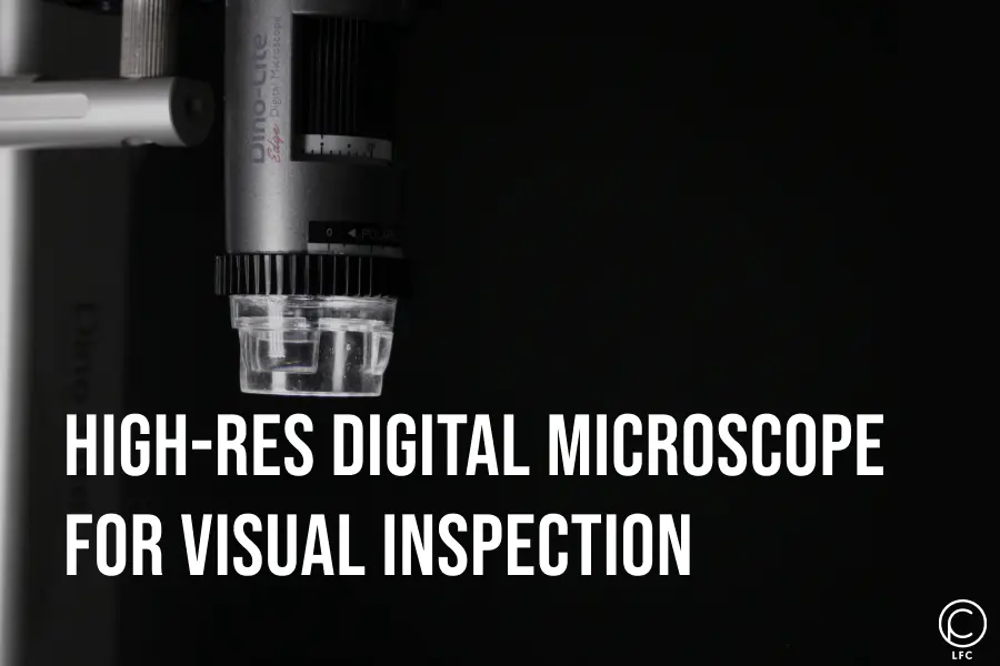
High-Resolution Digital Microscope for Visual Inspection
In digital microscopy, image quality and detail clarity are key factors determining a device's effectiveness. Dino-Lite has emerged as one of the leading solutions for high-quality visual inspection, combining advanced technology with ergonomic design to deliver impressive results across various fields of research and industry.
With its advanced features, Dino-Lite enables researchers and professionals to perform in-depth analysis on micro samples, enhancing work efficiency and accuracy of results. This microscope speeds up observation processes and paves the way for discoveries that can have a significant impact.
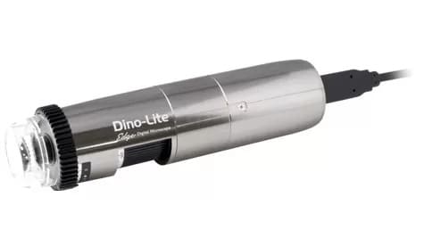
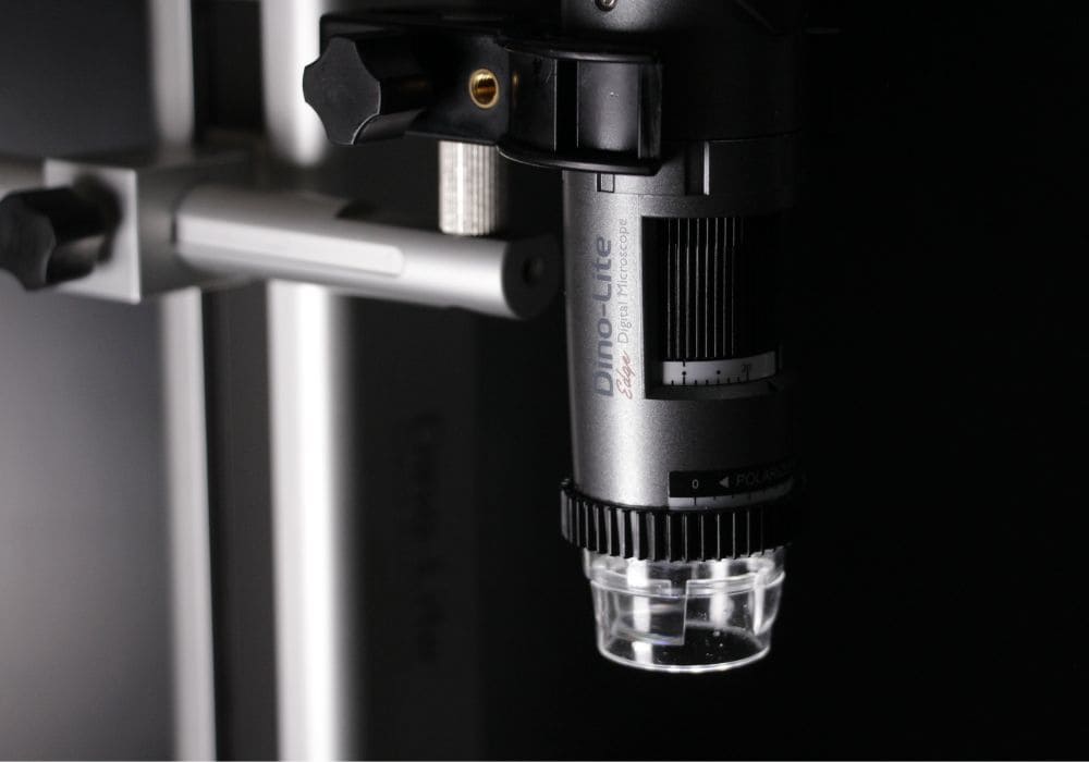
What is the Dino-Lite AM8917MZT?
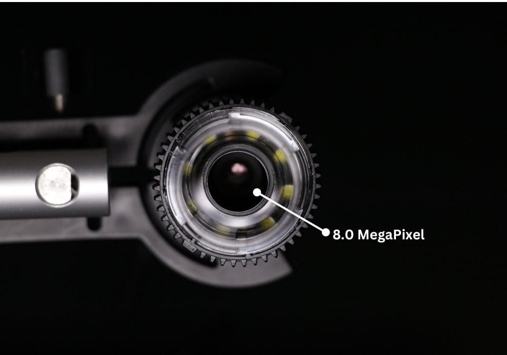
The Dino-Lite AM8917MZT digital microscope is a high-quality microscope with an 8MP resolution capable of capturing images of observed objects and producing 4K Ultra High-Resolution images with sharp, detailed clarity. Its anodized aluminum alloy construction protects against electromagnetic interference, ensuring durability and long-term use.
The Dino-Lite AM8917MZT digital microscope features technology that allows direct integration with a computer via a USB connection. This makes it easy for users to capture images and videos directly and access additional analysis features available in the software.
- Resolution: 8MP (3840 x 2160)
- Field of View (FOV): 16:9 Aspect Ratio
- Magnification Range: 10x - 220x
- Features: Integrated polarizer, DPQ, EDOF, EDR, AMR, and eFLC
- Software: DinoCapture 3.0 (New Version)
Advantages of the Dino-Lite AM8917MZT Digital Microscope
1. Portability: Its compact size makes it easy to carry anywhere.
2. Flexibility: It can be used in various lighting types and conditions.
3. Optical Quality: 8MP sensor that produces sharp, high-quality images with a 16:9 aspect ratio.
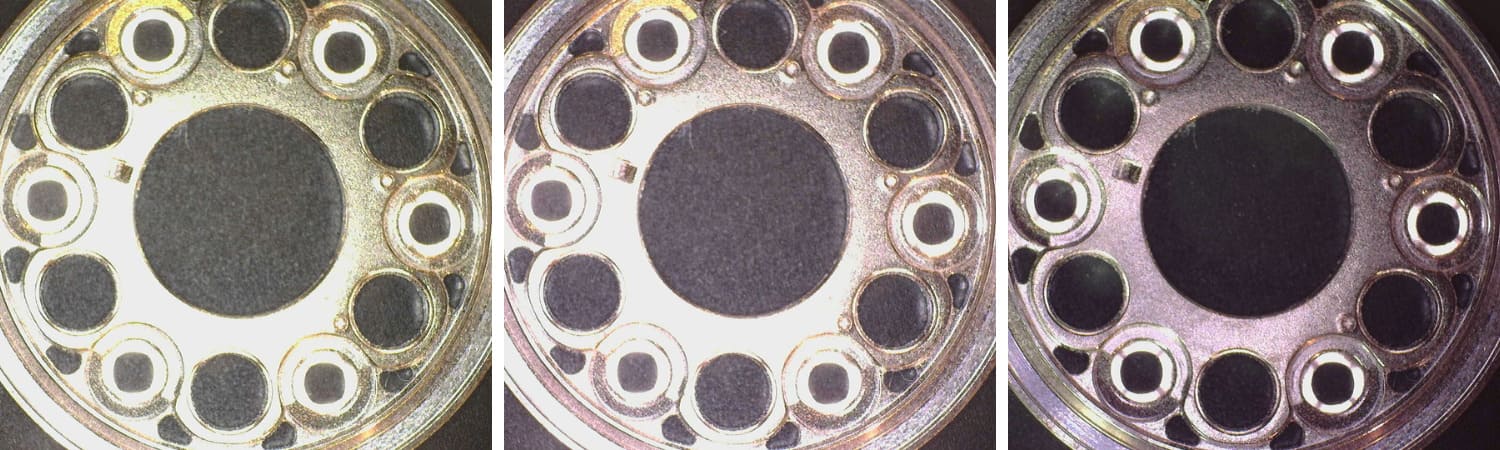
4. Light Control:
- eFLC: Flexible light intensity adjustment across two groups of LED quadrants.
- Polarizer: Removes unwanted glare from object surfaces.
5. Depth of Focus:
- DPQ: Precision focus control for accurate depth information.
- EDOF: Combines multiple images with different focus points to view rough surfaces.
- EDR: Displays details of dark or bright areas by merging images with different exposure levels.
6. Focus and Magnification:
- Auto/Manual Refocusing: Adjust focus with a click or mouse scroll.
- AMR: Automatically detects magnification power in real-time.
Applications of the Dino-Lite AM8917MZT Digital Microscope
With its exceptional capabilities, the Dino-Lite AM8917MZT digital microscope is highly suitable for various applications, including:
1. Quality Control (QC)
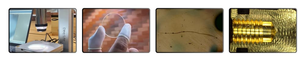
In QC, the Dino-Lite AM8917MZT enables users to measure, inspect surface quality, and detect product defects with detailed, high-quality images.
2. Education
 The Dino-Lite AM8917MZT digital microscope is a teaching aid that displays the structures of small objects in detail. It is highly effective for educational purposes and allows the analysis of various materials.
3. Electronics

The Dino-Lite AM8917MZT digital microscope makes it easy for users to examine electronic components such as PCBs, soldering, etc.
4. Healthcare
In healthcare, the Dino-Lite AM8917MZT enables professionals to examine and document biological samples such as skin, hair, and tissue with a high level of detail.
Digital Microscope Distributor in Singapore
LFC PTE LTD is the authorized distributor of Digital Microscopes in Singapore. Our expert team can assist you in selecting the ideal microscope to meet your specific requirements. Follow LFC PTE LTD on social media to stay updated on Dino-Lite products and other visual equipment. You can find us on Facebook, Instagram, YouTube, and LinkedIn.
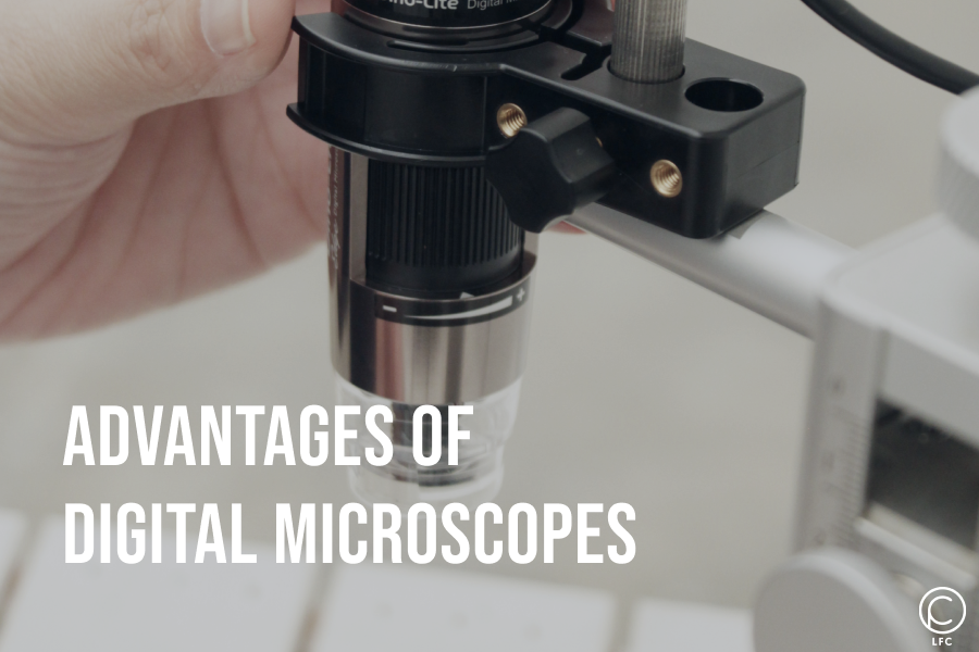
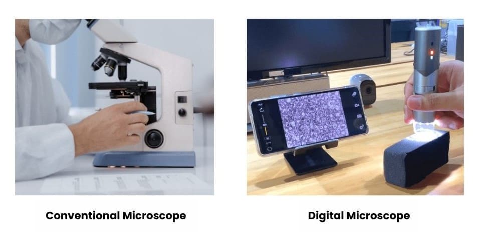
Advantages of Digital Microscopes Compared to Conventional Microscopes
What is a Conventional Microscope?
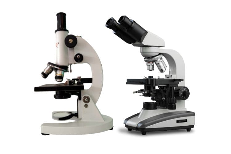
A conventional optical microscope magnifies objects using visible light and a lens system between the sample and the observer’s eye. The light is transmitted and passes through objective and ocular lenses to the observer's eye.
What is a Digital Microscope?

A digital microscope uses a digital camera instead of an eyepiece. Digital microscopes are generally handheld portable devices that can connect to a computer monitor, allowing real-time viewing of the results.
In a digital microscope, optics and a digital camera capture images displayed on a computer monitor. Some advanced features include image editing, 3D sample analysis, measurement, and data transfer.
Differences Between Conventional Optical Microscopes and Digital Microscopes
Digital microscopes perform the same tasks as optical microscopes but have many additional features. Some differences between digital and conventional optical microscopes include:
| Feature | Conventional Optical Microscope | Digital Microscope |
| Depth of Field | Higher magnification results in a shallower depth of field | The depth of field is 20 times more significant compared to traditional microscopes |
| Variable Angle Observation | The sample must be viewed directly from the above | Sampel dapat diamati dalam sudut 360 derajat |
| 3D Imaging | Produces 2D images with hard-to-determine surface characteristics | Produces entirely focused 3D images |
| Quantitative Data | No measurement or quantitative data features are available | Real-time measurement features available |
| Data Sharing | Not possible | Images can be recorded and saved directly |
Why Should You Use a Digital Microscope?
1. Ergonomics
A digital microscope is the right choice if you work in a lab with high sample throughput or need to use a microscope for hours. With a digital microscope, images can be displayed directly on a computer screen, allowing you to analyse them comfortably.
2. 360 Degree Observation
Digital microscopes offer variable angle capabilities, allowing 360-degree observation, so you don’t need to tilt your sample. Additionally, the time required for inspection is significantly reduced.
3. Fitur multi-lighting
Some digital microscopes come equipped with multi-lighting features to prevent glare issues when capturing images during observation.
4. Wide Range of Magnification
Digital microscopes offer a wide and varied magnification range, up to 900x, which allows users to analyse samples in depth and detail.
5. Integrated Archiving System
You can save images in different file formats after observing and selecting images. Some digital microscopes can be used for digital image analysis integrated with software.
Digital Microscope Distributor in Singapore
LFC PTE LTD is the authorised distributor of Digital Microscopes in Singapore. Our expert team can assist you in selecting the ideal microscope to meet your specific requirements. Follow LFC PTE LTD on social media to stay updated on Dino-Lite products and other visual equipment. You can find us on Facebook, Instagram, YouTube, and LinkedIn.

The Digital Microscope: A Modern Tool for Exploration
A digital microscope provides excellent observation results because it captures high-contrast images independent of the observer’s visual sharpness. What are the advantages and features of a digital microscope? Read on for a detailed explanation of digital microscopes.
What is a Digital Microscope?
A digital microscope is an advanced microscope equipped with a small digital camera for capturing images. The images viewed through the digital microscope can be projected onto a computer monitor and saved in computer files for future use. If the computer is connected to a digital printer, the images can also be printed.
Differences Between Digital and Optical Microscopes
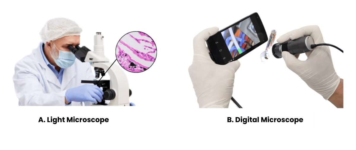 Optical microscopes were in use long before the development of digital microscopes. They require users to adjust lens combinations to achieve the desired magnification and focus, which can be challenging, especially for untrained users or those with impaired vision.
In contrast, digital microscopes are much easier to use, as users can simply view the image of the specimen on a monitor screen. The images produced are highly contrasted and clear, which is why many industries have now adopted digital microscopes.
Advantages of Digital Microscopes
Digital microscopes offer the following advantages:
- With a digital USB microscope, you can capture images and view them in 3D
- Images can be magnified and captured in real-time
- Images can be stored and transferred to a computer
- Versatile for various types of observations.
- Equipped with features that support high-quality imaging.
- Easy and quick to operate
Components of a Digital Microscope
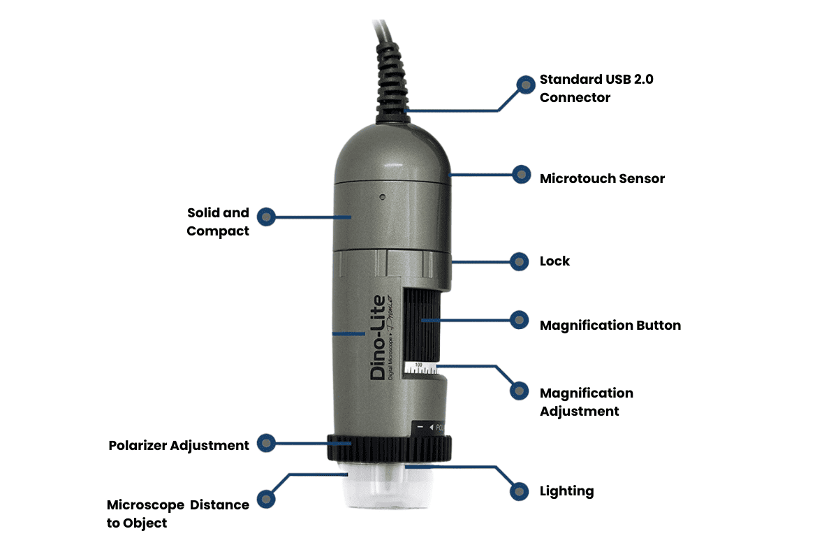 Digital microscopes consist of the following components:
- USB Connector: A USB cable connects the digital microscope to a computer.
- Microtouch Sensor: A touch screen used to capture images and control LEDs.
- Scroll Lock: Locks the the selected magnification level
- Magnification dial: Used to adjust the microscope’s magnification power
- Adjustable Magnification: Displays variable magnification options.
- Lighting: LED lights provide illumination during observations.
- Polarizer Adjustment: An optical filter that reduces glare or polarized light.
- Optical Camera: Captures images at specified resolutions and sizes.
Key Features of Digital Microscopes
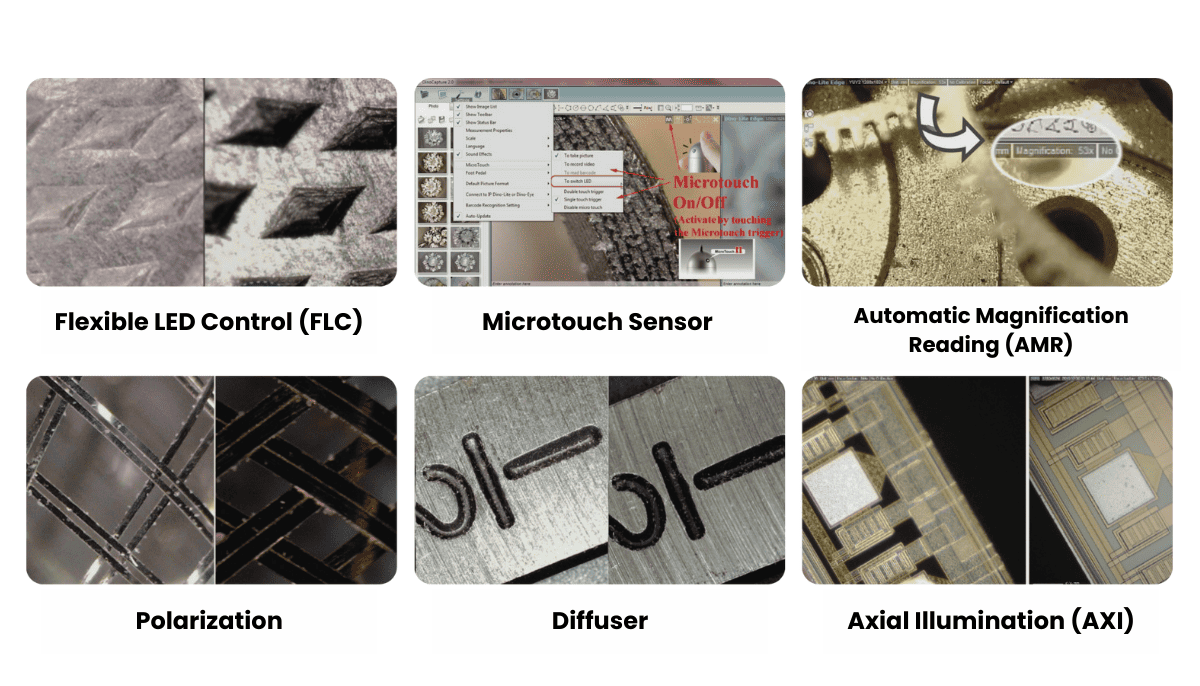 Some standard features of Dino-Lite digital microscopes include:
1. Flexible LED Control (FLC)
Adjust LED brightness and turn LEDs on/off by quadrant.
2. Microtouch Sensor
Trigger images are captured and controlled through DinoCapture (Windows) or DinoXcope (Mac OS).
3. Automatic Magnification Reading (AMR)
AMR is an automatic magnification reading feature that displays magnification through software. AMR is only available on specific Edge Series digital microscope models.
4. Polarization
Some models include a polarizer that can be turned on/off or adjusted for full or partial polarization.
5. Diffuser
A polyoxymethylene plastic insert that diffuses light to reduce glare.
6. Axial Illumination (AXI)
It provides axial or brightfield illumination, which is adjustable via software.
For detailed information on digital microscope features, visit our featured page.
Where Are Digital Microscopes Used?
Digital microscopes are used across many sectors, including:
1. Industrial Sector
Ideal for visual inspection, scratch detection, corrosion, and wear analysis in industries like textiles, metals, aerospace, and electronics.
2. Medical Sector
Used for medical examination, dermatology, and trichoscopy.
3. Jewel and Craft Sector
Essential for high-focus work on precious metals and gemstones.
4. Educational Sector
Due to its convenient use, it makes it easy for students to observe biological specimens.
5. Research and Development Sector
It enables researchers to study microscopic organisms and specimens with micron-level precision.
Check out our solution page to learn how to get the most out of your digital microscope.
Examples of Digital Microscopes
1. Dino-Lite EdgePLUS AM4917MZT

This model offers precise Z-axis height/shift measurement using enhanced EDOF technology, which is highly sought after by professionals.
2. Dino-Lite Edge AF4915ZT

This model is designed to meet diverse professional needs, and the optional WF-20 allows for wireless connection to PCs and phones.
3. Dino-Lite UV AM4113FVT

Equipped with UV LED lighting around the 400 nm spectrum, this model magnifies up to 200x, revealing detailed information on various objects.
4. Dino-Lite Basic AM2111

A USB digital microscope with simple features for capturing and storing images and videos, suitable for beginners and elementary education.
Digital Microscope Distributor in Singapore
LFC PTE LTD is the authorized distributor of Digital Microscopes in Singapore. Our expert team can assist you in selecting the ideal digital microscope to meet your specific requirements. Follow LFC PTE LTD on social media to stay updated on Dino-Lite products and other visual equipment. You can find us on Facebook, Instagram, YouTube, and LinkedIn.

How Professionals Use Dino-Lite Microscopes in Various Fields
Precision and clarity are paramount in the dynamic landscape of modern industries. Professionals across various fields rely on advanced tools to maintain high standards and achieve unparalleled accuracy. The Dino-Lite digital microscope has emerged as a versatile and indispensable device among these tools. Renowned for its portability, user-friendliness, and exceptional imaging capabilities, the Dino-Lite microscope has found applications in numerous sectors, including forensics, manufacturing, electronics, and quality control.
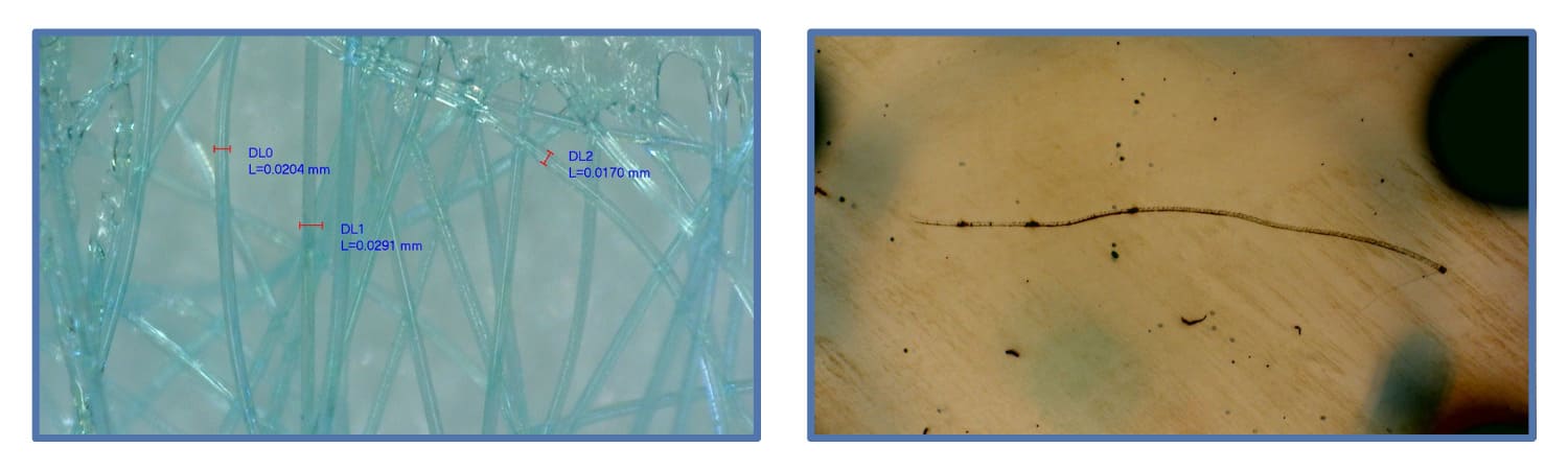
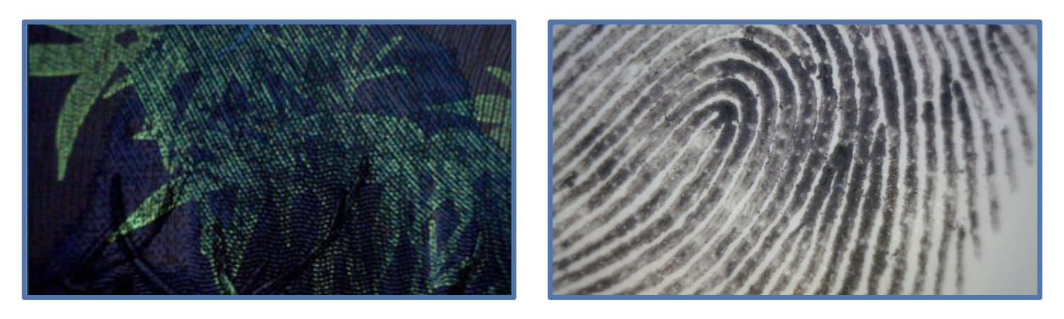
Forensics: Unveiling the Truth with Clarity

The Dino-Lite microscope is an essential tool in forensics, where every detail can be the difference between justice and injustice. Forensic scientists use these microscopes to meticulously examine evidence such as hair fibres, skin cells, and fabric threads. The high magnification and superior imaging quality of the Dino-Lite allow for the detailed analysis of minute samples that are often crucial in criminal investigations.
One notable application is in the analysis of counterfeit documents. Forensic document examiners employ Dino-Lite microscopes to scrutinise the fine details of suspected forgeries. The microscope’s ability to capture high-resolution images aids in identifying inconsistencies in ink patterns, paper fibres, and printing techniques. This meticulous examination is instrumental in verifying the authenticity of documents and can be pivotal in legal proceedings.
Additionally, Dino-Lite microscopes are utilised in crime scene investigations. Investigators can use these portable devices to examine trace evidence on-site, providing immediate insights without transporting samples to a lab. This capability not only speeds up investigations but also preserves the integrity of the evidence, ensuring that crucial details are not lost or contaminated during transit.
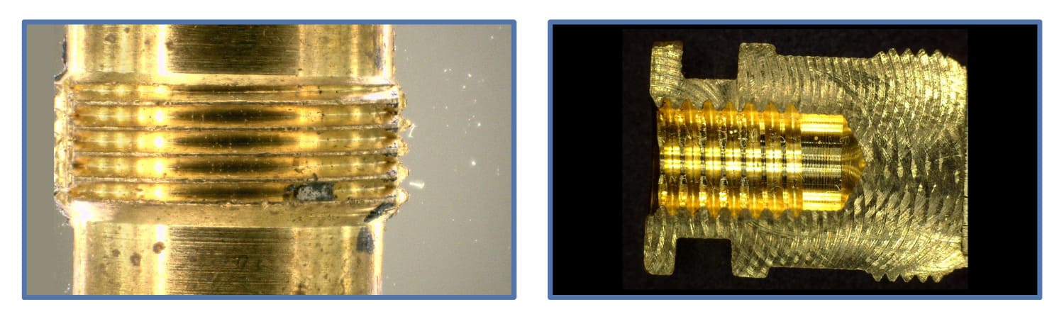
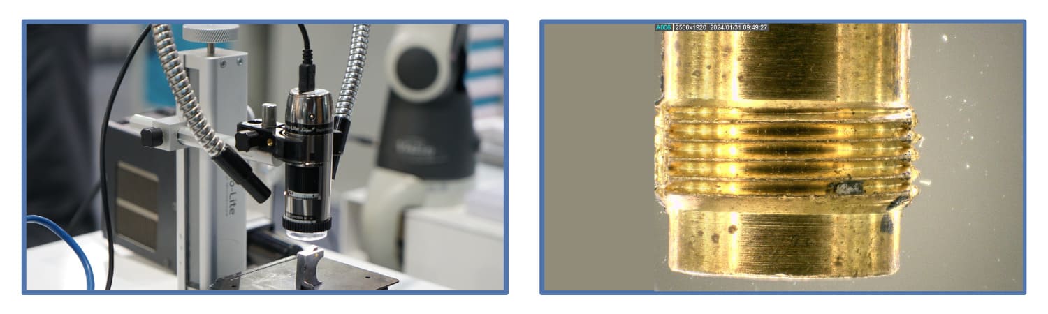
Manufacturing: Ensuring Precision and Quality

In the manufacturing sector, precision is synonymous with quality. Dino-Lite microscopes are crucial in ensuring that products meet stringent quality standards. From automotive components to medical devices, manufacturers use these microscopes to inspect parts for defects, measure dimensions, and verify compliance with specifications.
For instance, even the slightest imperfection can lead to significant malfunctions in electronic component production. Dino-Lite microscopes enable manufacturers to inspect solder joints, circuit boards, and microchips with exceptional clarity. The high magnification allows the detection of invisible defects to the naked eye, ensuring that only flawless components move forward in the production process.
Furthermore, Dino-Lite microscopes are invaluable in the quality control of precision-machined parts. Engineers and technicians use these devices to measure surface roughness, check for microscopic cracks, and ensure that parts adhere to exact specifications. The ability to capture detailed images and perform measurements directly on-screen enhances efficiency and accuracy in quality control processes.
Electronics: Enhancing Detail and Reliability
The electronics industry, characterised by rapid innovation and miniaturisation, demands tools to keep pace with its evolving needs. Dino-Lite microscopes have become indispensable in this field, allowing engineers and technicians to examine and troubleshoot complex electronic assemblies.
In PCB (printed circuit board) inspection, Dino-Lite microscopes identify manufacturing defects, such as solder bridges, component misalignments, and damaged traces. The microscopes’ high-resolution imaging and adjustable lighting options allow for a thorough examination of intricate PCB layouts. This detailed inspection is crucial for ensuring the reliability and performance of electronic devices.
Moreover, Dino-Lite microscopes are used to repair and rework electronic components. Technicians rely on these devices to inspect and repair small-scale components, such as microchips and connectors. The microscopes’ precision and versatility enable technicians to confidently perform delicate tasks, reducing the risk of further damage and improving the success rate of repairs.
Quality Control: Maintaining Standards Across Industries

Quality control is a critical aspect of any industry, and Dino-Lite microscopes have proven to be valuable assets in maintaining high standards. These microscopes are used in various quality control applications, from inspecting raw materials to verifying finished products.
For example, Dino-Lite microscopes are used in the pharmaceutical industry to inspect drug formulations and packaging materials. The microscopes’ ability to provide clear, magnified images aids in detecting contaminants, ensuring that products are safe for consumption. Additionally, in the food and beverage industry, Dino-Lite microscopes are used to examine food samples for foreign particles and quality issues, helping to uphold safety and quality standards.
Another significant application of Dino-Lite microscopes is in inspecting textiles and garments. Manufacturers use these devices to check for defects in fabric, such as weaving errors and surface irregularities. Dino-Lite microscopes' high magnification and imaging capabilities enable detailed inspection, ensuring that only high-quality textiles reach consumers.
Conclusion: A Tool for Precision and Excellence

The Dino-Lite microscope has become a versatile and essential tool across various industries. Its forensics, manufacturing, electronics, and quality control applications underscore its importance in modern professional practices. Dino-Lite microscopes empower professionals to achieve excellence in their respective fields by providing unparalleled clarity and precision. As industries evolve and demand higher standards, the Dino-Lite microscope will remain a cornerstone of accuracy and quality assurance.
In forensics, the ability to scrutinise evidence with unmatched detail aids in the pursuit of justice. In manufacturing, ensuring precision and quality translates to superior products. The electronics industry benefits from the microscope’s capability to enhance the reliability of intricate assemblies, while quality control across various sectors is elevated by its detailed inspection capabilities. The Dino-Lite microscope exemplifies how advanced technology can revolutionise practices, improve outcomes, and set new benchmarks for excellence in professional fields.
As technology advances, the potential applications of Dino-Lite microscopes are bound to expand further. Their role in enhancing precision, efficiency, and quality across industries highlights the transformative impact of innovative tools in the hands of skilled professionals. The Dino-Lite microscope is not just a tool; it is a testament to the power of technology in driving progress and achieving unparalleled standards of excellence.

The Advantages and Applications of Dino-Lite Edge Microscopes
Dino-Lite Edge is a sophisticated series of digital microscopes that provides high-quality imaging and advanced application features. These devices have gained popularity due to their portability, ease of use, and ability to deliver precise and detailed images. Unlike traditional microscopes, Dino-Lite Edge incorporates modern technology, making it a valuable tool in multiple industries. The series is known for its superior image quality, enhanced functionality, and versatility, making it a preferred choice for professionals and enthusiasts.
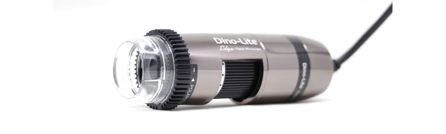
The Dino-Lite Edge series features a range of models, each tailored to meet specific needs and applications. These digital microscopes are equipped with state-of-the-art optics, adjustable lighting, and user-friendly software, allowing for detailed analysis and documentation. Whether used in laboratories, manufacturing, or fieldwork, Dino-Lite Edge offers a reliable microscopic examination and measurement solution.
Advantages of Dino-Lite Edge
1. Superior Image Quality
 One of the standout features of the Dino-Lite Edge series is its exceptional image quality. The microscopes are equipped with high-resolution sensors that capture clear and detailed images. The Edge series also incorporates advanced optics and adjustable polarisation, reducing glare and enhancing contrast, making it easier to observe fine details. This superior image quality is crucial in applications requiring precision, such as quality control, research, and medical diagnostics.
2. Portability and Ease of Use
 Dino-Lite Edge digital microscopes are designed with portability in mind. Their compact and lightweight design allows users to carry them easily and use them in various settings. Additionally, these devices are user-friendly, with intuitive controls and software that simplify capturing and analyzing images. The microscopes' USB connectivity ensures they can be easily connected to computers or other devices, enabling seamless data transfer and real-time viewing.
3. Versatility
Another significant advantage of the Dino-Lite Edge series is its versatility. These microscopes come with interchangeable caps and various magnification options, allowing users to customize the device to their needs. Whether examining electronic components, biological specimens, or industrial materials, the Dino-Lite Edge can adapt to various applications. The ability to switch between different magnifications and lighting conditions enhances the scope of analysis and ensures accurate results.
4. Advanced Software
Advanced software enhances the functionality of Dino-Lite Edge microscopes. The software offers measurement tools, image annotation, and the ability to capture time-lapse videos. Users can also calibrate their microscopes to ensure accurate measurements and documentation. The software's compatibility with various operating systems further adds to its convenience, making it accessible to many users.
5. Cost-Effective
Compared to traditional microscopes, Dino-Lite Edge digital microscopes are relatively cost-effective. They provide high-quality imaging and advanced features at a fraction of the cost of conventional microscopes. This affordability makes them attractive for educational institutions, small businesses, and professionals who require reliable microscopy solutions without a significant financial investment.
Applications of Dino-Lite Edge
1. Quality Control and Inspection
 In manufacturing and quality control, the Dino-Lite Edge series plays a crucial role in inspecting and ensuring the integrity of products. These microscopes can examine electronic components, machined parts, and other materials for defects or inconsistencies. The ability to capture high-resolution images and perform precise measurements helps identify issues early in the production process, reducing waste and improving overall product quality.
2. Medical and Clinical Applications
 The medical field benefits significantly from the advanced features of Dino-Lite Edge microscopes. These devices are used in dermatology, pathology, and other medical specialties to examine tissues, cells, and other biological samples. The Dino-Lite Edge's portability allows for its use in clinics, laboratories, and even fieldwork, providing healthcare professionals with a versatile tool for diagnosis and research.
3. Education and Research
 Educational institutions and research laboratories utilize Dino-Lite Edge microscopes for teaching and scientific investigation. The ease of use and affordability make these devices ideal for students learning about microscopy and researchers conducting detailed studies. The ability to capture and share images and videos enhances the learning experience and facilitates collaboration in research projects.
4. Forensics and Law Enforcement
 Forensic experts and law enforcement agencies use Dino-Lite Edge microscopes for crime scene investigation and evidence analysis. These microscopes can reveal minute details crucial for forensic analysis, such as examining fibers, documents, and trace evidence. Dino-Lite Edge's portability and advanced imaging capabilities make it an invaluable tool in forensic science, aiding in the accurate and efficient analysis of evidence.
5. Art and Conservation
Dino-Lite Edge microscopes examine and document artworks and artifacts in art restoration and conservation. These devices help conservators analyze the materials and techniques used in artworks, identify areas of damage, and plan restoration efforts. The high-resolution imaging and ability to capture detailed images are essential for preserving the integrity and authenticity of historical and cultural objects.
6. Agriculture and Environmental Science
Dino-Lite Edge microscopes are also employed in agriculture and environmental science to study plants, soil, and other natural materials. These devices help researchers and professionals examine plant structures, identify pests and diseases, and monitor environmental changes. The Dino-Lite Edge's portability allows for field studies, making it a valuable tool for on-site analysis and research.
Conclusion
 The Dino-Lite Edge series of digital microscopes offers numerous advantages and applications across various industries. Its superior image quality, portability, versatility, advanced software, and cost-effectiveness make it a preferred choice for professionals and enthusiasts. Whether used in quality control, medical diagnostics, education, forensics, art conservation, or environmental science, the Dino-Lite Edge provides a reliable and efficient microscopic examination and analysis solution. As technology advances, the capabilities and applications of Dino-Lite Edge microscopes are likely to expand, further solidifying their position as essential tools in numerous fields.
Advanced Imaging Techniques with Dino-Lite: Tips for High-Quality Microscopy
Microscopy has long been a cornerstone of scientific research, allowing us to explore the intricate details of the microscopic world. With technological advancements, microscopy has evolved significantly, providing researchers with powerful tools to observe and analyze samples with unprecedented clarity and precision. Among these tools, Dino-Lite stands out as a leader in the field, offering versatile and high-quality imaging solutions for a wide range of applications. In this article, we'll explore some advanced imaging techniques with Dino-Lite and provide tips for achieving high-quality microscopy results.
Understanding Dino-Lite Microscopes
 Dino-Lite microscopes are renowned for their compact design, ease of use, and exceptional image quality. They utilize advanced optics and cutting-edge digital imaging technology to deliver clear, detailed images of microscopic specimens. Dino-Lite offers a variety of models tailored to different applications, including biological research, industrial inspection, forensics, and more. These microscopes feature adjustable magnification levels, adjustable LED illumination, and a range of accessories to enhance imaging capabilities.
Advanced Imaging Techniques with Dino-Lite
 In the realm of microscopy, Dino-Lite stands as a beacon of innovation and excellence, offering cutting-edge imaging solutions that empower researchers, educators, and enthusiasts to explore the microscopic world with unparalleled clarity and precision. With its compact design, user-friendly interface, and advanced imaging capabilities, Dino-Lite has revolutionized how we perceive and analyze the tiny wonders that lie beyond the threshold of the naked eye. In this section, we delve into advanced imaging techniques with Dino-Lite, uncovering the secrets of the microscopic universe through a lens of unprecedented sophistication and insight.
-
High-Resolution Imaging
At the heart of Dino-Lite's imaging prowess lies its ability to capture high-resolution images of microscopic specimens with breathtaking detail and fidelity. Equipped with state-of-the-art sensors and optics, Dino-Lite microscopes deliver images that rival the clarity and resolution of traditional laboratory-grade microscopes, allowing users to discern the subtlest features of their samples with precision and accuracy. Whether you're examining biological specimens, geological formations, or industrial materials, Dino-Lite's high-resolution imaging capabilities offer a window into a world of exquisite complexity and beauty.
-
3D Imaging and Depth Profiling
Beyond traditional 2D imaging, Dino-Lite microscopes can capture three-dimensional images of microscopic specimens, providing valuable insights into their topographical structure and morphology. By utilizing advanced imaging techniques such as structured light illumination and confocal microscopy, Dino-Lite enables users to visualize the three-dimensional architecture of their samples with unprecedented clarity and depth. From intricate cellular landscapes to rugged geological formations, 3D imaging with Dino-Lite unveils the hidden dimensions of the microscopic universe, offering a new perspective on the complicated interplay of form and function.
-
Fluorescence Imaging
Fluorescence microscopy is a powerful technique for visualizing specific molecules and structures within biological specimens, and Dino-Lite microscopes are equipped to harness their full potential. Dino-Lite enables users to visualize fluorescently labeled molecules and structures with exquisite specificity and sensitivity by incorporating fluorescence filters and light sources into their imaging systems. From tracking cellular processes' dynamics to unraveling molecular interactions' mysteries, fluorescence imaging with Dino-Lite opens new avenues for exploration in biological research and discovery.
-
Time-Lapse Imaging and Live Cell Imaging
Dynamic processes unfold in real-time within the microscopic world, and Dino-Lite microscopes can capture these phenomena through time-lapse imaging and live cell imaging. By capturing sequential images at defined intervals, Dino-Lite enables users to observe and analyze dynamic processes such as cell division, migration, and signaling with unparalleled temporal resolution. Whether you're unraveling the mysteries of embryonic development or studying the behavior of living cells in response to external stimuli, time-lapse imaging with Dino-Lite provides a window into the dynamic rhythms of life at the microscopic scale.
-
Spectral Imaging and Hyperspectral Analysis
Spectral imaging is a powerful technique for analyzing the spectral characteristics of microscopic specimens, and Dino-Lite microscopes offer advanced capabilities in this domain. By capturing images at multiple wavelengths across the electromagnetic spectrum, Dino-Lite enables users to perform hyperspectral analysis, revealing valuable information about their samples' chemical composition and molecular structure. Whether identifying specific biomolecules within cells or characterizing the composition of geological minerals, spectral imaging with Dino-Lite offers many opportunities for spectroscopic analysis and discovery.
Tips for High-Quality Microscopy with Dino-Lite
Microscopy, powered by Dino-Lite technology, opens a window to the intricate world of the microscopic, offering researchers and enthusiasts a glimpse into realms unseen by the naked eye. To harness the full potential of your Dino-Lite microscope and unlock the mysteries held within your samples, here are some advanced tips for achieving impeccable microscopy results:
-
Optimize Illumination
As mentioned earlier, proper illumination is the cornerstone of high-quality microscopy. With Dino-Lite microscopes, fine-tune the LED illumination to match the specific characteristics of your sample. Avoid extreme overexposure or underexposure, as these can obscure details and compromise contrast, leading to subpar images that fail to do justice to your samples' intricacies.
-
Use Polarization
 Harness the power of polarization many Dino-Lite models offer to combat glare and elevate contrast in your images. By experimenting with different polarization settings, you can minimize unwanted reflections and unveil the delicate nuances hidden within transparent or reflective samples, revealing a wealth of detail that might otherwise go unnoticed.
-
Adjust Focus and Magnification
 Precision is paramount when it comes to focus in microscopy. Utilize the fine focus knob on your Dino-Lite microscope to meticulously adjust focus, ensuring that your images are sharp and crisp. Furthermore, explore the range of magnification levels available to capture every intricate detail of your samples, from the grandeur of macroscopic structures to the subtleties of cellular morphology.
-
Explore Different Lighting Modes
 Dino-Lite microscopes offer many lighting modes, each tailored to illuminate specific types of samples optimally. From traditional brightfield illumination to the dramatic contrasts of darkfield and the nuanced revelations of differential interference contrast (DIC), experiment with different lighting modes to uncover the full spectrum of details in your samples, choosing the mode that best complements your specific application.
-
Utilize Measurement and Annotation Tools
 Beyond mere observation, leverage Dino-Lite software's measurement and annotation tools to delve deeper into your microscopy findings. Accurately measure dimensions, annotate vital features, and generate comprehensive reports for further analysis and sharing, transforming your microscopy endeavors into a rich tapestry of quantifiable data and insightful observations.
-
Calibrate Your Microscope
Adhere to regular calibration procedures for your Dino-Lite microscope to maintain the integrity of your microscopy data. Follow the manufacturer's guidelines meticulously, conducting calibration checks at regular intervals to ensure precise measurements and consistent imaging results, laying a solid foundation for your scientific endeavors.
-
Consider Environmental Factors
Environmental conditions can significantly impact microscope performance and image quality. To mitigate potential disruptions, ensure that your Dino-Lite microscope is in a stable environment with controlled temperature, humidity, and minimal vibrations, providing a conducive setting for optimal imaging results.
-
Practice Proper Sample Preparation
Last but certainly not least, meticulous sample preparation is the bedrock upon which high-quality microscopy is built. Adhere to established protocols for sample fixation, staining, and mounting, ensuring that your samples are meticulously prepared to withstand the scrutiny of Dino-Lite microscopy. Investing time and effort into proper sample preparation will pave the way for insightful observations and groundbreaking discoveries.

Educational Applications of the Dino-Lite Digital Microscope
In the ever-evolving landscape of education, technological advancements continue to revolutionize how students learn and engage with subject matter. One such innovation that has gained traction in recent years is the Dino-Lite digital microscope.
Initially developed for industrial and professional use, this powerful tool has found its way into classrooms and educational institutions worldwide, opening up a new realm of possibilities for hands-on learning in the sciences and beyond.
Unveiling the Microscopic Universe
 The Dino-Lite digital microscope offers a window into the microscopic universe, allowing students to explore the intricate details of objects at magnifications ranging from 10x to 220x and beyond. Equipped with high-quality optics and advanced imaging technology, these microscopes deliver stunning clarity and resolution, enabling users to observe everything from biological specimens to geological samples with unprecedented precision. With features such as adjustable LED illumination and polarizing filters, students can customize their observations to suit their specific needs and objectives.
Bridging Theory and Practice
 One of the most significant advantages of incorporating the Dino-Lite digital microscope into educational settings is its ability to bridge the gap between theoretical concepts and real-world observations. Instead of relying solely on textbooks and diagrams, students can now directly observe cellular structures, microorganisms, and other phenomena, gaining a deeper understanding of their subjects. Through interactive exploration, students can test hypotheses, make observations, and draw conclusions, engaging in authentic scientific inquiry.
Enhancing STEM Education
In science, technology, engineering, and mathematics (STEM), the Dino-Lite digital microscope is a valuable tool for enhancing learning outcomes and fostering curiosity. Immersing students in hands-on exploration encourages critical thinking, problem-solving, and inquiry-based learning. Whether examining the anatomy of a leaf or investigating the behavior of microorganisms, students develop essential scientific skills while gaining a newfound appreciation for the natural world. Furthermore, the microscope's compatibility with various digital platforms and software applications enables students to analyze and manipulate data, reinforcing their understanding of STEM concepts.
Supporting Multidisciplinary Studies

Beyond the sciences, the applications of the Dino-Lite digital microscope extend into various other disciplines, including art, history, and archaeology. For instance, art students can analyze pigment particles and brush strokes in famous paintings, while history enthusiasts can examine ancient artifacts and fossils in intricate detail. By integrating microscopy into multidisciplinary studies, educators can enrich the learning experience and inspire interdisciplinary thinking among students. Furthermore, the microscope's ability to capture high-resolution images and videos facilitates documentation and analysis, allowing students to present their findings creatively and engagingly.
Engaging Learners of All Ages
One of the most remarkable aspects of the Dino-Lite digital microscope is its versatility, making it suitable for learners of all ages and abilities. Whether in primary, high, or university, students can explore the microscopic world at their own pace, guided by educators who facilitate discovery and inquiry. The user-friendly interface and intuitive controls make it accessible to individuals with diverse learning styles and special needs, ensuring that no one is left behind in pursuing knowledge. Through collaborative projects and peer-to-peer learning experiences, students develop essential teamwork and communication skills, preparing them for success in future academic and professional endeavors.
Fostering Collaboration and Exploration
 In addition to its educational benefits, the Dino-Lite digital microscope facilitates collaboration and exploration both inside and outside the classroom. With features like live video streaming and image capture, students can share their discoveries with peers and experts, sparking discussions and collaborative research projects. Furthermore, the portability of these microscopes allows for field trips and outdoor expeditions, where students can conduct real-time observations in various natural environments. By connecting with scientists, researchers, and educators worldwide, students gain insights into cutting-edge research and applications, inspiring them to pursue further studies and careers in STEM.
Looking Ahead
As technology advances, the educational applications of the Dino-Lite digital microscope are poised to expand even further. With ongoing developments in imaging technology, connectivity, and software integration, educators can expect new opportunities to enhance the learning experience and inspire the next generation of scientists, researchers, and innovators. Whether through virtual reality simulations, remote collaboration tools, or artificial intelligence applications, the microscope will continue to play a pivotal role in shaping the future of education and scientific discovery.
Digital Microscope Distributor in Singapore
LFC PTE LTD is the official distributor of digital microscopes in Singapore. Please contact our team to discuss your company’s need for a digital microscope.
You can also visit our Facebook, Instagram, YouTube, and LinkedIn pages to get the latest updates about other industrial equipment.
