
Over the past eight years, the Dino-Lite digital microscope has helped users around the world increase their productivity in a wide range of mundane to mission-critical applications. Due to its superior image quality, compact form factor, and ease-of-use, this product has quickly risen to a dominant position in the marketplace. From its initial success, the product line has expanded to include hundreds of models ( and accessories) to fulfill specific customer needs. As a result, the Dino-Lite microscope is now an indispensable tool in multiple market sectors, such as academics, manufacturing, quality control, healthcare, and education.
1. Art Conservation & Restoration
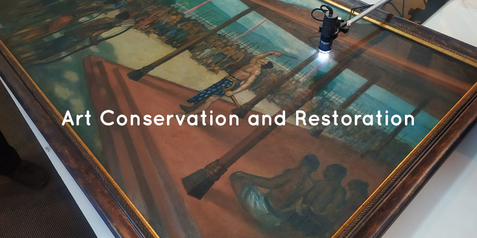
Art conservation is both an art and a science. The object of interest ranges from sculptures, ceramics, paper scroll (books, rolls, etc.), textiles and even architecture. Some of these could be performed in the lab, while others can only be done onsite. A microscope is a necessary tool in art conservation and restoration. They are used in non-destructive analysis and characterisation tests of the piece. Hence, the Dino-Lite digital microscope being small, light and portable is a great help for anyone in this field of work.
Dino-Lite digital microscope is a good support tool for art conservationists to restore and conserve objects and works of artists from the past. The portable digital microscope is held comfortably in your hand. It connects to a laptop via USB cable. The magnified image then appears on the screen through the DinoCapture 2.0 software. The alternative will be to look through an eyepiece of a stereo microscope. The Dino-Lite digital microscopes comes with high magnification and built-in LED lights to illuminate the object being examined. It is a portable microscope that will be a useful visual aid to see details that cannot be seen by the naked eye. The small and light form factor makes it useful for analysing large sized artworks which can be difficult to reach with the traditional stereo microscope.
When used to inspect sculptures, ceramics or architecture, the digital microscope can also help to identify the different types of stone, such as marble or limestone. It can also be used to analyse the components in mortars. With the DinoCapture 2.0 software, users will also be able to easily take pictures or videos for documentation. Other analysis tools available on the software includes measurements of line, circle, diameter, radius, angle, centre distance and etc. As such, it is a neat tool for measuring the size of the sand granules in mortar. It can also be used as a diagnostic tool for identifying the source of the stone’s deterioration, helping to identify salts, pollution and biological growth. It can even locate historic tool marks and past conservation treatments. When looking at a stain at a higher magnification, as a user, you would be able to differentiate if the stains come from pollutions or fungus for example.
The special lighting models with UV or infrared lights can help the user to view hidden details found on the art piece. For instance, the AM4115-FKT comes with 780 nm infrared LED lighting and records high quality images at 1.3 Megapixels. This model can also magnify objects up to 200x. It is ideal to be used to analyse chemical and physical properties of paper. On the other hand, the AF4515T-FVW comes with a 400 nm UV light source. The UV light source is a good aid for revealing historical moisture and mould damage to a paper. These are not always visible to the naked eye. Areas of the paper which were previously in contact with water, where size and the products which cause fluorescence have been removed, appear darker under UV light. All this information can then be used to aid the decision making process of forming a suitable conservation treatment.
Art restoration and conservation is a process that needs to be carried out very carefully. Art restorers would normally need to clean the art objects with great care so as not to damage the last original paint coat. They would need to differentiate between dirt, excess paint and original paint layer. They would also need to determine the authenticity of the items by looking at the object structure. With microscopes that can magnify items at 200x magnification, Dino-Lite can help to analyse in detail visually the different materials used to produce the art.
Dino-Lite being used to check a painting and projected on a wide TV screen.
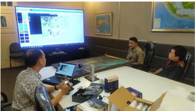
The user will also find Dino-Lite USB microscope’s ability to capture images and videos, which can be annotated, to be very useful for documenting historical art objects. The user would also have the flexibility to select the medium they wish to view the art with. Dino-Lite digital microscopes can display images on TV screens, PC monitors, laptops and even Android devices. Art professionals can easily observe the work without any contact with the microscope’s ability to view items at a distance with the long working distance models of Dino-Lite.
Some might argue that without digital microscopes, conservators in the field can still turn to handheld loupes for analysis. However, the magnification powers of such loupes can only go up to about 30x. At the end of the day, the samples viewed will still need to be taken to a laboratory for analysis. Whereas the Dino-Lite USB microscope offers magnification of up to 250x. Analysis can be done immediately when you are on field. In the case of stone conservators, after looking at the stone with the Dino-Lite digital microscope, they can take samples, for example, of each kind of biological growth and bring them back to the lab for further analysis. The information can then be used to decide treatment plans for the long-term care and conservation of the stone monuments and buildings.
The Dino-Lite digital microscopes have time and again proven to itself to be a cost-effective tool. Coupled with the free DinoCapture 2.0 software, it will greatly help to speed up the analytical process in conservation studies of many different types of materials.
References
1. Nichols, E. (2016, December 14). Under the microscope – preliminary investigations of the changi archive conservation research project. Retrieved April 14, 2021, from https://specialcollections-blog.lib.cam.ac.uk/?p=13554
2. Lee, C. (2020, November 02). Conservation tools: The usb digital microscope. Retrieved April 14, 2021, from https://blogs.getty.edu/iris/conservation-tools-the-usb-digital-microscope/
