
Practical platform for a routine check up and observation. Dino-Lite microscope provide one of the most important platform in studying biological sciences towards medical, trichoscopy and dermatology observation. It allows practitioner and patients to conveniently venture into the world of the cell, as well as discover the fascinating world of microscopic feature and draw a diagnosis precisely.
1. Dermatology - Scalp / Hair
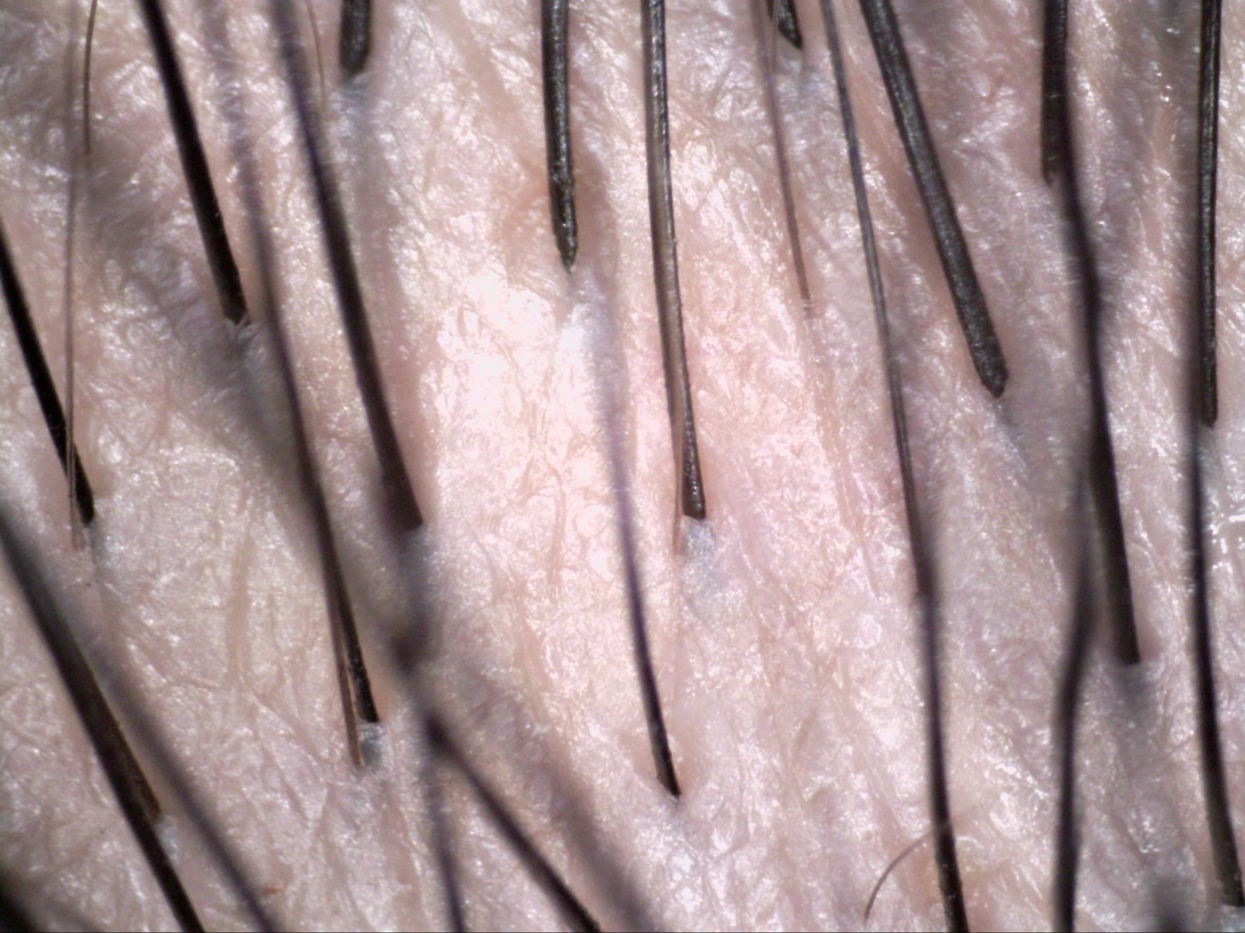
Here are some recommended Dino-Lite digital microscopes for dermatology-scalp/hair:
2. Nail Capillary
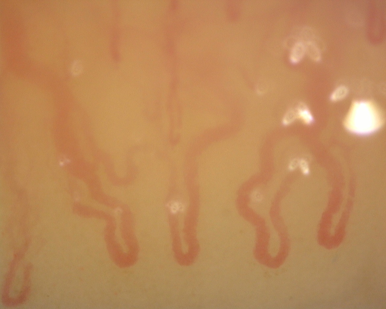
A capillary is a small blood vessel from 5-10 micrometres in diameter. They are the smallest blood vessels in the body: they convey blood between the arterioles and venules. Physician could diagnose various kinds pf disease by checking up the nail capillary.
Dino-Lite Nail Microcirculation Scope has a magnification of 500x and can examine the capillary with magnified clarity and digitally record the image for diagnostic and other purposes.
Here are some Dino-Lite Microscope recommended for observing nail capillary :
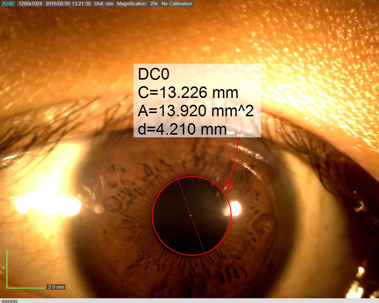
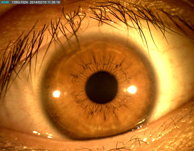
3. Ophthalmology

Ophthalmology is the field of medicine that deals with the anatomy, functions, and diseases of the eye. This includes the diagnosis and treatment of disorders of the eye. An ophthalmologist is a physician who specializes in ophthalmology. The credentials include a degree in medicine, followed by an additional four to five years of residency training in ophthalmology. Residency training programs for ophthalmology may require a one-year internship with training in internal medicine, pediatrics, or general surgery. Additional specialty training (or fellowship) may be sought in a particular aspect of eye pathology. Ophthalmologists are allowed to prescribe medications to treat eye diseases, implement laser therapy, and perform surgery when needed. Ophthalmologists may participate in academic research on the diagnosis and treatment of eye disorders.
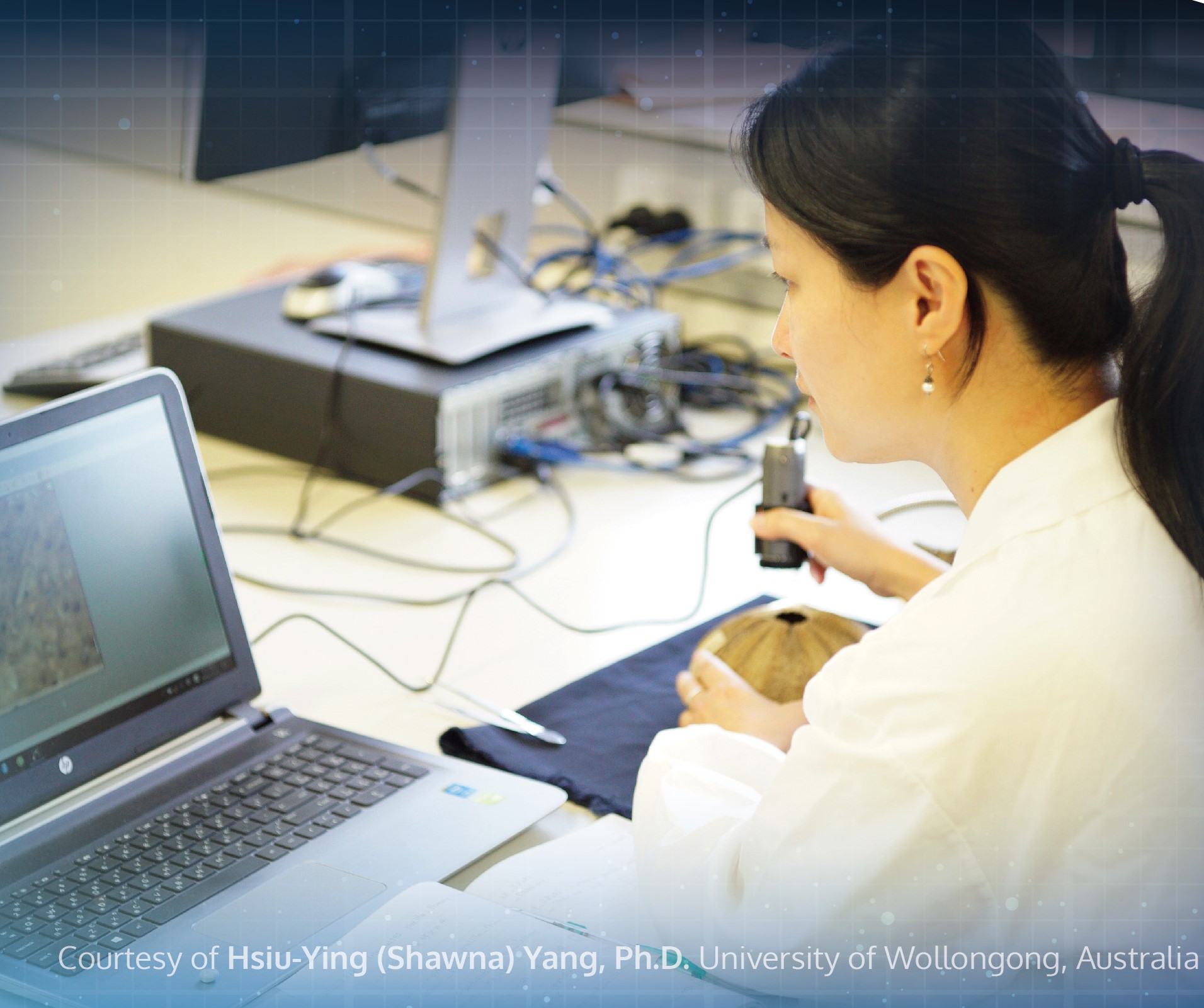
Improved View
Dino-Lite digital microscopes can help ophthalmologists when you are doing iris examinations. The AM4113-RUT is one of the many models by Dino-Lite that is suitable for such work. It is a USB microscope which is used by connecting it to a computer. The main benefit of using digital microscopes is improved view. As a user, you would be able to examine the iris through a large screen monitor instead of hunching over patients. Ophthalmologists typically must hunch to get a close examination of the patients’ iris through oculars. The AM4113-RUT enables you to sit comfortably throughout the day as you are examining patients one after another. Extended periods of bending over oculars to examine multiple patients in a day may create a cumulative strain on your back. Over time, it can be damaging to your back and cervical spine. The traditional approach takes a lot of wear and tear on your bodies. Studies show that between 30% and 50% of ophthalmologist end up with cervical neck pain or herniated discs due to the way you must position your bodies to view the eye with a traditional microscope.
Ease of Use
The AM4113-RUT is easy to use. It is very much like the other Dino-Lite models where you merely use a single scroll to magnify and focus all at once. Your movements and hand placement may be different than using oculars. Most ophthalmologists are used to resting your head against the microscope as you look through the oculars. With the AM4113-RUT, you would no longer need to rest your head against the oculars of the microscope. It is a minor change that one can easily adapt to. Rest assured that the learning curve of using such a digital microscope is not steep. As a user, you would not require a long time to get accustomed to the Dino-Lite as it is as simple as viewing an image on the screen.
Data Collection
USB models of Dino-Lite digital microscopes are used with the DinoCapture 2.0 software. The application exists to support ease of documentation of your iris examination. You would be able to take pictures or videos by using the MicroTouch function. On the AM4113-RUT, there is a touch sensitive trigger for taking pictures and videos. Upon a single click, a picture is taken and automatically saved on your computer. All recent videos and images taken can be viewed on the left-hand bar of the DinoCapture 2.0 software. You can view the information seamlessly without interruptions during the examination. This is also a neat software for organizing and filing of patients’ iris examination images. It is easy to transfer images and videos from the DinoCapture 2.0 as they are saved to a designated folder in your computer. As you begin to use the digital microscope, you can get used to taking advantage of its capabilities. You will find an increase in efficiency as a benefit upon switching to the digital microscope. It will also greatly help you if you are seeking to file data in a “paperless” manner.

Communal Viewing
Anyone in the examination room — assistants, technicians, medical students — can watch the examinations on a monitor. As a result, it will help them to understand the patients’ situation better. If we compare it to the use oculars for examinations, only the person viewing through the microscope can get the complete picture. The others in the room will not be able to look at a monitor. The ability to see exactly what the doctor is seeing is an incredible advantage for teamwork overall. Everyone can better anticipate what is happening during the examination. Further, it also serves as a tool for resident education, fellow education, and medical student education because everyone is seeing the same thing at the same time.
Increased Safety
In the COVID-19 pandemic, digital microscopes offer an additional barrier of safety for doctors and patients. Being able to examine patients through the monitors, and not through the oculars, allows the ophthalmologist to wear a full-face shield. There is a great safety advantage to having the ability to sit back and not hover directly over the patient’s face during examination. The ability to cover your face and eyes with a face shield is an important layer of protection, for both patients and doctors amid the pandemic. Information captured is also directly saved into the computer. You would not need to handle information on written copies. You would be able to access the images again by going through them in the computer. As a result, you would be less prone to error. Data that is captured goes straight from the digital microscope directly to the computer. It increases efficiency and is better for patient safety. When examining multiple patients, you would want to do it with as little room for error as possible. Digital microscopes close that gap quite a bit.
Here is the USB Dino-Lite Digital Microscope recommended for ophthalmology use:
